Figure 1: Representational image showing a typical ultrasound sensor
Bats are wonderful creatures. Blind of sight and yet such precise vision that he could distinguish between a moth and a broken leaf even when he was flying at full speed. There is no doubt that vision is sharper than ours and far beyond the human ability to see, but it is certainly not beyond our understanding. Ultrasonic ranging is the technique used by bats and many other creatures in the animal kingdom for navigation purposes. In an attempt to imitate the ways of nature to gain an advantage over everything, we humans have not only understood it, but have also successfully imitated some of these manifestations and made the most of their potential.
History

Figure 2: Graphic image displaying ultraviolet sensor history
The story dates back to 1790, when Lazzaro Spallanzani first discovered that bats maneuvered in flight using hearing instead of vision. Jean-Daniel Colladon, in 1826, discovered ultrasonography using an underwater bell, successfully and accurately determining the speed of sound in water. After that, study and research work in this field proceeded slowly until 1881, when Pierre Curie's discovery set the stage for modern ultrasound transducers. He discovered the relationship between electrical voltage and pressure in crystalline material. The unfortunate accident of the Titanic sparked a rigorous interest in this field, and as a result Paul Langevin invented the hydrophone for detecting icebergs. It was the first ultrasonic transducer. The hydrophone could send and receive low-frequency sound waves and was later used in detecting submarines in the First World War.
In parallel with SONAR, medical research also began to take an interest in ultrasound. In the late 1930s, Dr. Karl Dussik used a technique called hyperphonography, which recorded echoes of ultrasonic waves on sensitive paper. This technique was used to produce ultrasound images of the brain to help detect tumors and marked the birth of ultrasound imaging. After that, many scientists like Ian Donald, Douglas Howry, Joseph Holmes, John Wild and John Reid improved the various aspects of ultrasonic sensors in the medical field which enabled the diagnosis of stomach cancer, ovarian cysts, detection of twin pregnancies, tumors, etc. . The industry wasted no time jumping on the bandwagon either and soon developed techniques such as ultrasonic welding and non-destructive testing in the early 1960s.
Working
How do ultrasonic sensors work?
Ultrasonic sensors are devices that use electromechanical energy transformation, with mechanical energy in the form of ultrasonic waves, to measure the distance from the sensor to the target object. Ultrasonic waves are longitudinal mechanical waves that travel as a succession of compressions and rarefactions along the direction of propagation of the wave through the medium. Any sound wave above the human hearing range of 20,000 Hz is called ultrasound. Depending on the type of application, the frequency range has been broadly categorized as shown in the figure below:

Figure 3: Graphical figure of sound frequency ranges
When ultrasonic waves fall on an object, the diffuse reflection of energy occurs at a wide solid angle that can reach 180 degrees. Thus, some fraction of the incident energy is reflected back to the transducer in the form of echoes and is detected. The distance to the object (L) can then be calculated from the speed of the ultrasonic waves (v) in the medium by the relationship
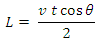

Figure 4: Diagram showing how ultrasound waves work
Where 't' is the time it takes for the wave to return to the sensor and '  'is the angle between the horizontal and the path taken as shown in the figure. If the object is moving, instruments based on Doppler shift will be used. Get all the details about the internal structure and working of an ultrasonic sensor in Insight-How Ultrasonic Sensors Work.
'is the angle between the horizontal and the path taken as shown in the figure. If the object is moving, instruments based on Doppler shift will be used. Get all the details about the internal structure and working of an ultrasonic sensor in Insight-How Ultrasonic Sensors Work.
Generation of ultrasonic waves
Generating Ultrasonic Waves
For the generation of such mechanical waves, the movement of some surface such as a diaphragm is necessary, which can then induce movement in the medium in front of it in the form of compression and rarefaction. Piezoelectric materials operating in motor mode and magnetostrictive materials have been widely used to generate ultrasonic waves in the frequency ranges of 1-20 MHz and 20-40 kHz, respectively. The sensors employ piezoelectric ceramic transducers that flex when an electrical signal is applied to them. These are connected to an electronic oscillator whose output generates the oscillating voltages at the required frequency. Materials such as Lead Zirconate Titanate are popular piezoelectric materials used in medical ultrasound imaging. For best results, the frequency of the applied oscillations must be equal to the natural frequency of the ceramic, which readily produces oscillations through resonance. Offers maximum sensitivity and efficiency when operated at resonance.
Piezoelectricity, being a reversible phenomenon, produces electrical voltages when ultrasonic waves reflect back from the target and collide with the ceramic structure. In this way, a transducer can function both as a transmitter and as a receiver in pulsed mode. When continuous measurement of distances is required, separate transducers can be used for transmission and reception. Sensors, when used in industry, are generally employed in assemblies that can be mechanical assemblies that consist of oscillating or rotating sensors, or electronic assemblies that can be linear, curved or phased. To visualize the output of an ultrasonic sensor, displays of different types are used, the format of which depends on the type of transducer set used and the function. A sectoral field of view is produced by mechanical arrays and curved and phased electronic arrays, while a linear field is generated by linear arrays. Display modes can be linear graphical plotting with amplitude on the y-axis and time on the x-axis, called Amplitude mode or A-mode, or intensity-modulated B-sweeps, where the brightness of a point indicates the amplitude of the reflected waves. Other modes include M mode, Doppler (D) mode, etc.
The parameterization of these sensors is generally done by monitoring the reflected and transmitted signals of the lateral and axial movement of the transducer, keeping the target fixed in a specific medium (water in general). The sound beam diverges quickly, so care is taken to ensure that the transducer produces the smallest possible beams. The narrower the beam pattern, the more sensitive the sensor. However, the possible angle between the transducer and the surface increases with the beam width. Beam patterns of the type shown below are observed:

Figure 5: Graphic image showing profile of axial and transverse beams
The parameters on which the performance of an ultrasonic sensor is measured include bandwidth, attenuation, dynamic range, and resolution such as grayscale, axial and lateral resolution. Other parameters are nominal frequency, peak frequency, bandwidth center frequency, pulse width, sensitivity and signal-to-noise ratio (SNR).
Importance and problems
Importance of Ultrasonic Sensors
There are a variety of sensors based on other physical transduction principles, such as optical range-finding sensors and also microwave-based devices. So, why should one use ultrasonic transducers in the first place, given that the speed of sound is much slower than the speed of electromagnetic waves? The answer is in the question itself. Because devices based on EM waves are very fast. Being slower than EM waves, the time taken by ultrasonic waves is much longer than that of the latter and, therefore, their measurement can be done more easily and less expensively. As they are based on sound waves and not EM waves, they would work in places where the latter would not.
For example, in the case of detecting bright objects and measuring liquid levels or high-brightness environments, light-based sensors would suffer greatly due to the transmittance of the target or the translucency of the propagation medium. Ultrasonic devices based on sound propagation would remain largely unchanged. They also work well in humid environments, where optical beams can suffer from refraction from water droplets in the environment. Due to range and accuracy, ultrasonic sensors can fall between two EM wave-based sensors, infrared rangefinders on the low end and LIDARs on the high end. Not as accurate or long-distance as LIDARs, ultrasonic rangefinders fare better than IR rangefinders, which are highly susceptible to environmental conditions and require recalibration when the environment changes. Additionally, these devices offer advantages in medical imaging compared to MRI or X-ray examinations due to their low cost and portability. No harmful effects of ultrasonic waves at the intensity levels used have been detected in contrast to X-ray or radioactivity-based methods and are particularly suitable for soft tissue imaging.
Problems and concerns
However, ultrasonic sensors are not free from all problems either. The speed of sound in a medium increases as the temperature of the medium increases. So, even when the target remains in the same location, it may now appear as if it has moved to a location closer to the sensor. Air currents due to various reasons can disturb the wave path, which can lead to “Missed Detection” or wrong measurement.
Acoustic noises, such as high-pitched sounds created due to hissing or hissing from valves and pneumatic devices at a frequency close to the operating frequency, can interfere with the sensor output. Electrical noise also affects sensor performance. These can generate artifacts that are not a true representation of the photographed object. Just as vision begins to become blurred when the distance from the object to the eye becomes too small for the eyes to see it, ultrasonic devices also have a “dead zone” where the sensor cannot make measurements reliably. This happens due to a phenomenon called ringing, which is the continuous vibration of the transducer after the pulse is emitted. Thus, when the distance is very small, the transducer has not yet stopped to be able to differentiate between the vibration due to the incident radiation or the oscillation of the electrical excitation. The dangers of ultrasonic waves are also well founded. If the intensity is too high, it can cause heating of human tissues and cause ruptures in people exposed to it. Ethical issues such as fetal identification and resulting abortions in the medical field are also a widespread concern.
Forms
The applications of ultrasonic sensors can be classified based on the property they exploit. They can be summarized as:
|
Domain
|
Parameter
|
Forms
|
|
Time
|
Flight block, speed
|
Density, Thickness, Flaw Detection, Anisotropy, Robotics, Remote Sensing etc.
|
|
Mitigation
|
Fluctuations in reflected and transmitted signals
|
Defect characterization, microstructures, interface analysis
|
|
Frequency
|
Ultrasonic Spectroscopy
|
Microstructure, granulometry, porosity, phase analysis.
|
|
Image
|
Mapping time of flight, speed and attenuation in Raster C-Scan or SARs
|
Images of surface and internal defects, density, speed, 2D and 3D images.
|
Research has been carried out to overcome the problems of ultrasonic sensors, particularly in medical imaging, where it is known as ultrasound. Artifacts from ultrasonic sensors such as Acoustic Shadowing and Acoustic Enhancement are being explored to characterize tissues that allow differentiation between solid and cystic tissues. The industry has also reaped the benefits of ultrasonic sensors in applications such as plastic welding, jewelry cleaning, remote sensing and telemetry, assisted parking systems, etc. Robotics is known for using ultrasonic rangefinders as a favorite tool for distance measurement and mapping. Even the fashion industry is using ultrasonic sensors in hairstyles, such as hair extension implants.

Figure 6: Diagram showing flaw detection using ultrasonic sensors
Application and Future
Future
Non-destructive testing and flaw detection uses ultrasonic waves in various modes such as Longitudinal mode (L wave) and Shear mode (S wave) to detect flaws in materials. With advances in Science, new materials are being developed that offer greater performance at lower voltages, such as capacitive micromachined ultrasonic transducers (CMUTs), which should have greater bandwidth and greater potential for integration with electronic circuits.
These devices provide non-invasive measurements for detecting problems in all types of materials, whether living tissue or non-living manufactured products. With a healthy track record of being able to detect many problems that would otherwise leave doctors stunned and the problem untreated, ultrasonic sensors offer a lot of promise even in the times to come. With the environmental and psychological effects of EM radiation exposure rigorously put under the scanner, ultrasonic applications are expected to prosper and offer a substantial alternative to contemporary technologies.